


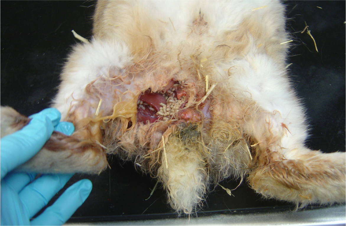
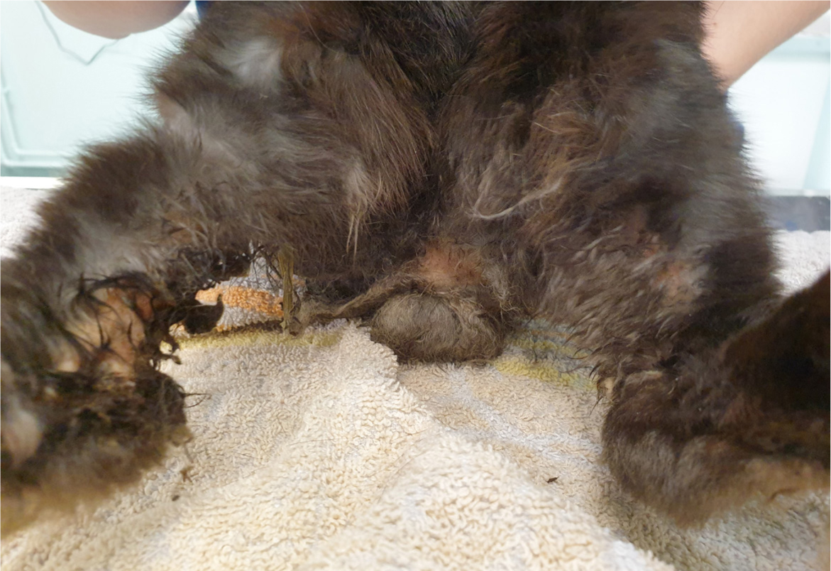
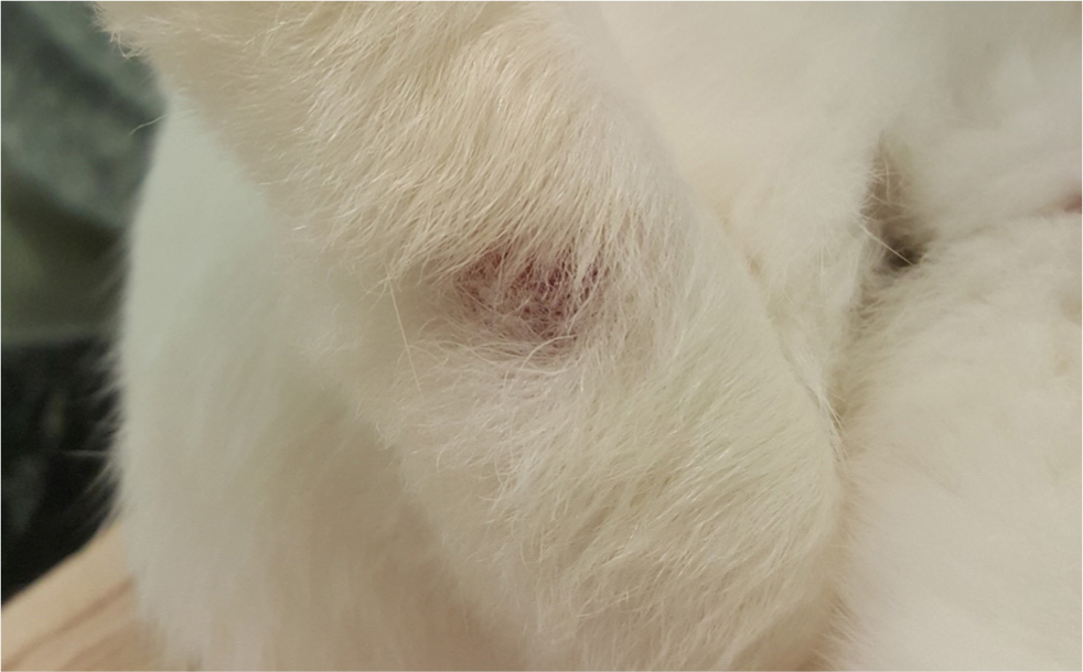
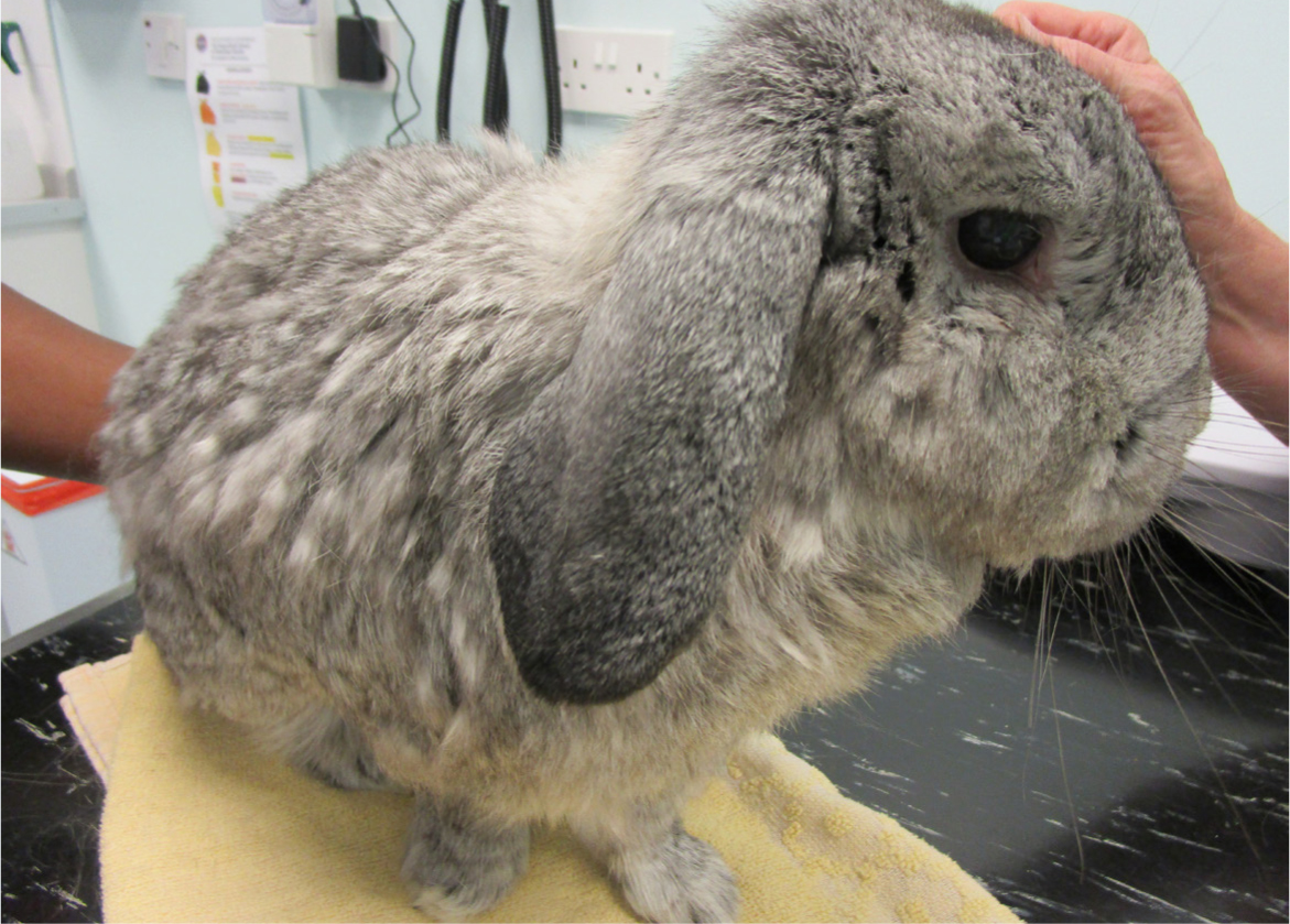
Osteoarthritis is defined as a disorder of articular joints, and involves deterioration of articular cartilage, osteophyte production and remodelling of bone in response to chronic damage to cartilage (Hunter et al, 2014). The recognised consequences of osteoarthritis in the diarthrodial joints are pain and disability (Fox, 2016). Osteoarthritis is the number one cause of chronic pain in dogs (Fox, 2016), and management of osteoarthritic pain is an important component of animal welfare. In rabbits, while pain caused by osteoarthritis is a significant factor in itself, secondary associated issues detrimentally impact on welfare. Rabbits unable to perform caecotrophy as a result of arthritic pain are susceptible to myiasis because of caecotroph accumulation in perineal skin (Figure 1). Urine scald may develop in rabbits that cannot position appropriately to urinate (Keeble, 2014) (Figure 2). An inflamed and infected perineum leads to development of urethritis, followed by cystitis caused by ascending infection (Varga, 2014). Reduced locomotion from pain predisposes to ulcerative pododermatitis, an extremely painful disease that is difficult to treat once established (Varga, 2014) (Figure 3). Rabbits are generally stoic and are known to hide signs of pain (McBride, 2014); consequently, chronic issues such as osteoarthritis may go unnoticed until advanced.
Although many studies investigate experimentally induced appendicular osteoarthritis in rabbits (Bouchgua et al, 2009; Dong et al, 2013; McCoy, 2015; Riester et al, 2017), few investigate naturally occurring osteoarthritis in client-owned rabbits under veterinary care (Arzi et al, 2011; Mäkitaipale et al, 2015). Increased understanding of risk factors for osteoarthritis in rabbits will allow preventative measures and management to be implemented against this welfare-threatening disease. Therefore, the aim of this study was to better understand this disease by reporting the prevalence and risk factors of computed tomography changes of osteoarthritis in pet rabbits. The hypothesis was that prevalence will increase with increasing age and weight, and that clinical detection will be low as a result of the prey status of rabbits and their ability to hide pain.
Materials and methods
Inclusion criteria
Domestic rabbits of any breed that had been admitted to the study hospital over a 4-year period (December 2017–October 2021) for a full body computed tomography scan for any reason were included in the study. If any individual rabbit had more than one computed tomography scan performed, only the first scan was included. Comorbidities were not recorded.
Computed tomography report evaluation
The study used only computed tomography reports recorded on ProVet Cloud, all of which had been written by a member of a dedicated diagnostic imaging team and cross checked by a specialist senior diagnostic imager at the time of the scan. Computed tomography reports for included scans were read, and presence of ‘osteoarthritis’ or ‘degenerative joint disease’, as well as the joint(s) involved were recorded using Microsoft Excel (v16.0). The joints of interest were the appendicular joints. The sacroiliac joint was included because of the involvement of the ilium, which is part of the appendicular skeleton (Anderson et al, 2023). The corresponding consultation and clinical examination were used to record information about the individual rabbit at the time of the computed tomography scan: the age in days, the weight in kilograms and the body condition score on a 5-point scale (UK Pet Food, 2022). The breed, sex and neuter status at the time of the scan were also recorded. The clinical examination notes that had been recorded for the computed tomography consultation were evaluated for description of findings associated with the presence of osteoarthritis. The key findings of interest were a description of ‘stiffness’, ‘reduced range of motion’ and ‘resistance/pain on manipulation of the limbs or joints’. The presence of these, or similar phrases, was used to decide if the condition was clinically apparent at the time of examination in rabbits that had computed tomography findings consistent with osteoarthritis, and was recorded as a ‘yes’ or a ‘no’. Rabbits that did not have any computed tomography findings consistent with osteoarthritis were marked as ‘not applicable’ in this category and excluded from the analysis of the ‘clinically apparent’ vs ‘non-apparent’ groups.
Statistical analysis
All statistical analyses were run using Minitab 20 Statistical Software. A chi-squared test was performed to evaluate association between sex and presence of osteoarthritis. The same test was run to evaluate association between neuter status and osteoarthritis in both the male and the female group. A chi-squared test was used to determine association between breed and presence of osteoarthritis. Breed data were present for 250 of 311 rabbits; 18 different breeds were present. Breeds with two individuals or fewer were eliminated from the analysis (Silver Fox, Rex, Polish, Plush Lop, Unspecified Lop, Harlequin and English Angora). The remaining breeds were placed in eight groups: Crossbreed, Continental Giant, English, Dutch, Netherland Dwarf, Lion Head, Giant Lop breeds (English Lop and French Lop), and Small Lop breeds (Dwarf Lop, Holland Lop, Mini Lop and Mini Lion Lop). When the expected cell counts were generated, 7/16 (44%) cells had expected values >5, meaning that the data sample was too small for accurate analysis using a chi-squared test (McHugh, 2013), and thus, no conclusion was drawn.
A Mann–Whitney test was run to assess a difference in body condition score in rabbits with and without evidence of osteoarthritis. The weight subset data were graphically assessed for normality; the data was not normally distributed (P<0.005), likely because of large variation between rabbits of different breeds. Therefore, a non-parametric test was used. Using the Minitab software, a Mann–Whitney test was not possible because the data was arranged with both groups in one column. For this reason, a Kruskal–Wallace test was run.
The data for age were recorded in days. For ease of interpretation, each value was divided by 365 before analysis to generate a value in years. The subset data were not normally distributed according to a graphical assessment (P<0.005), and therefore, a non-parametric test was used. The same software limitation was present as in the weight analysis, precluding use of a Mann–Whitney test. A Kruskal–Wallace test was run. For all tests, a P value of less than 0.05 was considered statistically significant.
Results
A total of 311 computed tomography reports were assessed. Out of 311 individual rabbit reports, 61 (19.6%) described the presence of osteoarthritis in at least one joint or joint pair. Of the rabbits with osteoarthritis, 33/61 (54.1%) had more than one joint type affected and 17/61 (27.9%) had three or more joint types affected (Figure 4). The most frequently affected joint was the elbow (44 rabbits; 14.1% of all rabbits), followed by the stifle (37 rabbits; 11.9% of all rabbits) (Table 1; Figure 5). Osteoarthritis was clinically evident in 9 out of 61 (14.8%) of rabbits, based on comments in the clinical exam notes on the day of the scan.
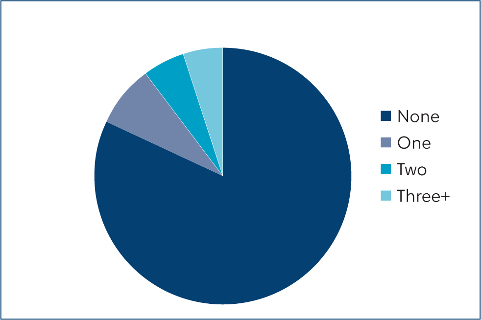
Table 1. Frequency of joints affected by osteoarthritis
| Joint affected | Number of rabbits with osteoarthritis in this joint type |
|---|---|
| Carpal | 8 |
| Elbow | 44 |
| Shoulder | 7 |
| Tarsal | 12 |
| Stifle | 37 |
| Coxofemoral | 5 |
| Sacroiliac | 1 |

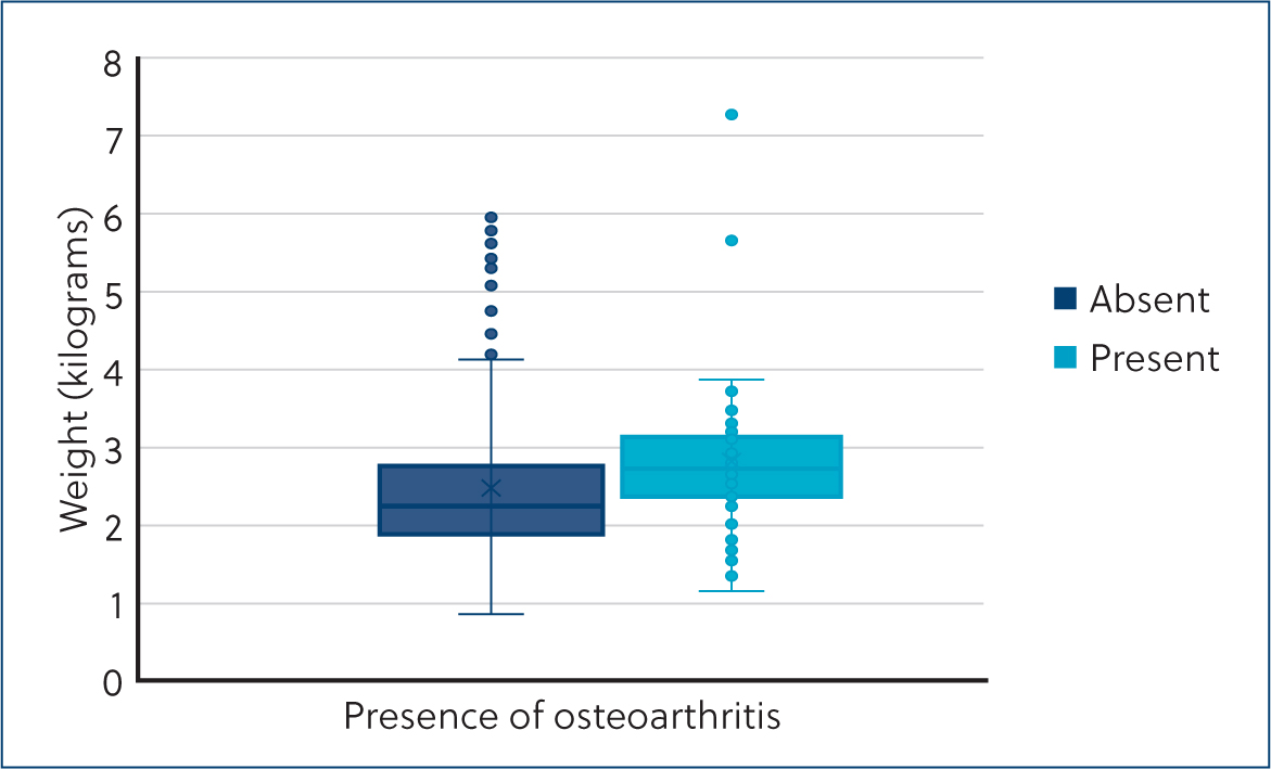
A Mann–Whitney test showed that in this sample, rabbits with osteoarthritis (median=2.5) and those without (median=2.5) did not have significantly different body condition scores (difference=0; 95% confidence interval -0.5–0.5; W-value=2171.0; p=0.791). The weight of rabbits included in the study ranged from 0.75 kg to 7.17 kg, with an average mean weight of 2.44 kg. A Kruskal-Wallis test showed that there was a statistically significant difference (H-value=10.71; degrees of freedom=1; p=0.001) in kilograms of weight between rabbits that did have evidence of osteoarthritis (median=2.60) and rabbits that did not have evidence of osteoarthritis (median=2.25) (Figure 6).
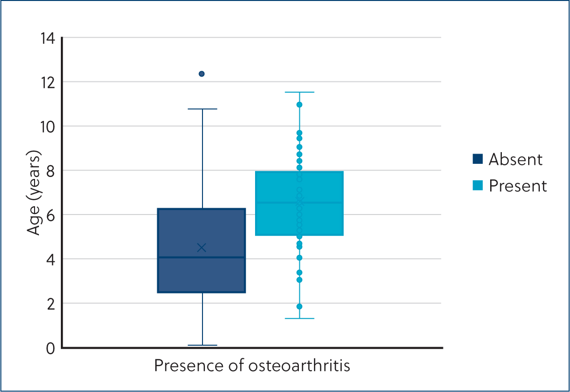
The age of rabbits included in the study ranged from 3 months to 12 years, with an average mean age of 5 years. A Kruskal–Wallis test showed that there was a statistically significant difference (H-value=32.44; degrees of freedom=1; P=0.000) in years of age between rabbits that did have evidence of osteoarthritis (median= 6.52) and rabbits that did not have evidence of osteoarthritis (median=4.01) (Figure 7).
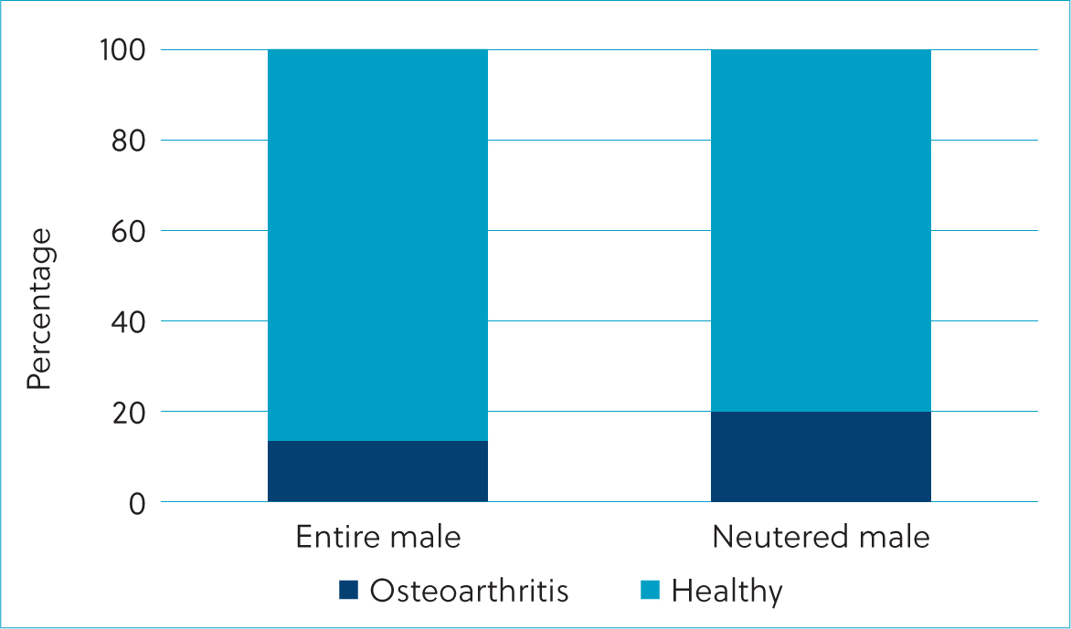
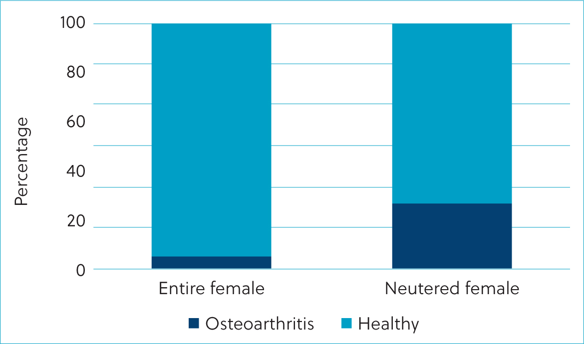
Of the rabbits included in the study, 131 were female and 180 were male. Of the female rabbits, 96 were neutered and of the male rabbits, 122 were neutered. Based on the results of a chisquared test, there was no association between presence of osteoarthritis and sex regardless of neuter status (chi-squared value (χ2)=0.442, degrees of freedom=1, P=0.506). In male rabbits, there was no association between presence of osteoarthritis and neuter status (χ2=1.226, degrees of freedom=1, P=0.268) (Figure 8). In female rabbits, there was a statistically significant association between presence of osteoarthritis and neuter status (χ2=8.467, degrees of freedom=1, P=0.004) (Figure 9). Therefore, neutered females had a higher prevalence of osteoarthritis than entire female rabbits. No conclusion was drawn concerning breed and osteoarthritis because the sample size was insufficient.
Discussion
In the population of this study, the prevalence of osteoarthritic changes on computed tomography was approximately 19.6%. A previous study that investigated naturally occurring osteoarthritis in rabbits under veterinary care found a prevalence of 40.2%, based on retrospective radiographical assessment of 189 rabbits (Arzi et al, 2011). A health survey looking at 167 apparently healthy pet rabbits found radiographical changes consistent with osteoarthritis in the distal limbs (carpus, elbow, stifle and hock) of 4/167 (2.4%) of rabbits (Mäkitaipale et al, 2015). Therefore, there is a large discrepancy in the estimated prevalence of osteoarthritis in pet rabbits. The current study used data from a single centre and only included rabbits that had indication for a computed tomography scan; therefore, it is unclear whether the prevalence observed can be extrapolated for any other population.
The most common joints affected in the study population were the elbow (14.1% of all rabbits) and the stifle (11.9%) (Figure 10). This is somewhat at odds with the findings of Arzi et al (2011), who found the most frequently affected joints on radiographic assessment to be the hip (26.1%) and the stifle (20.9%). It is postulated that computed tomography imaging is more accurate for assessment of the small rabbit elbow joint than radiography, as a detailed three-dimensional image is obtained. Differences in positioning and presence of normal overlying bone on radiographs of this joint could lead to false negatives on radiographic reporting for osteoarthritis. Additionally, radiography only detects moderate to advanced lesions associated with osteoarthritis and will not detect early subtle pathology (Arzi et al, 2011; Ley et al, 2021). Therefore, computed tomography is recommended for early disease detection (Arzi et al, 2011; Ley et al, 2021).
No conclusion could be made on the relationship between breed and presence of osteoarthritis. The analysis was limited by the low sample size and the large number of breed categories. A lower prevalence of osteoarthritis has previously been found in dwarf breeds (Arzi et al, 2011), and giant breeds are thought to be more prone to developing osteoarthritis (Varga, 2014).
Sex regardless of neuter status, and neuter status in males also had no apparent relationship with presence of osteoarthritic changes on computed tomography. However, a relationship was found between neuter status in females and presence of osteoarthritis, where neutered females had a greater presence of changes. This finding might have been confounded by greater age or weight in neutered females vs entire females in the sample population. Neutered animals have a tendency to put on more weight, which could have affected the development of osteoarthritis. It would have been interesting to have evaluated this further by looking at the incidence of osteoarthritis according to neuter status in all rabbits vs non-neutered status.
Neuter status has been observed as a risk factor in dogs of both sexes, but the same confounding factors exist in many studies (Anderson et al, 2020). In humans, prevalence in both sexes is equal until the age of 55 years, after which women are more frequently affected (Hunter et al, 2014). The proposed mechanism is that postmenopausal women lose a protective element from oestrogen. This concept is supported by the fact that hormonal therapy has been proven to delay the onset of arthritis (Hunter et al, 2014). Therefore, the general indication is that sex hormones may have protective function on the joints. Nevertheless, the argument for neutering female rabbits is extremely compelling, considering the high incidence of uterine adenocarcinoma in entire rabbits (Greene, 1941; Varga, 2014). Based on the findings of this study, the recommendation to neuter female rabbits does not change, but clinicians should consider a potentially higher prevalence of osteoarthritis in female neutered rabbits when in clinical practice.
Obesity or high body condition score is strongly related to osteoarthritis in dogs (Sanderson, 2012) and in humans (Garstang and Stitik, 2006; Martel-Pelletier et al, 2016). In contrast, the current study found that there was no significant relationship between body condition score and presence of osteoarthritic changes on computed tomography. One thing to consider is that the present study looked at body condition at a single point in time, at the time of the computed tomography scan, and did not consider a potential history of obesity, which may have been important, especially in aged rabbits that may have lost condition as a result of underlying concurrent health concerns such as renal or dental disease (Figure 11).
The current study found a significant relationship between higher weight and computed tomography evidence of appendicular osteoarthritis. This finding is consistent with risk factors for osteoarthritis in other species including dogs (Anderson et al, 2020) and humans (Martel-Pelletier et al, 2016). This conclusion also agrees with findings in Arzi et al's (2011) study.
The present study also found a positive relationship between greater age and evidence of osteoarthritis on a computed tomography scan. This finding is not unexpected, given that age is a significant and well-described risk factor for osteoarthritis in both dogs (Anderson et al, 2018; 2020) and humans (Hunter et al, 2014; Martel-Pelletier et al, 2016).
Clinical examination findings consistent with osteoarthritis were noted in only 14.8% of rabbits that had changes on the computed tomography scan. This suggests that approximately 85% of cases with a computed tomography diagnosis of osteoarthritis are not clinically apparent, which is a welfare concern. Several possibilities might explain this low level of clinically apparent disease: poor correlation of computed tomography findings with overt clinical disease, hiding of clinical signs because rabbits are a prey species or poor recognition of pain and gait changes associated with osteoarthritis by owners and veterinary staff. One study looking at stifle osteoarthritis in dogs suggested that radiographical evidence of osteoarthritis poorly relates to loss of limb function (Gordon et al, 2003). However, human studies found a consistent association in the knee and inconsistent association in the hip (Kinds et al, 2011). It is possible that computed tomography evidence of osteoarthritis is variably related to clinical evidence, with mild changes that may not be causing pain being more easily detectable on computed tomography imaging (Ley et al, 2021). However, given the range of movement of the rabbit elbow, stifle and hip joint during locomotion, as well as the great forces applied to these joints when pushing off the ground and landing during hopping movements, as compared with dogs, it would seem likely that loss of range of joint movement as a result of arthritic changes would impact on gait in the rabbit.
A comparative study using kinetic gait analysis in healthy vs osteoarthritic dogs has been published (Brønniche Møller Nielsen et al, 2020); however, this study concluded that the overlap performance was insufficient and therefore, general use of this method for diagnostic purposes in dogs with osteoarthritis could not be advocated. While kinetic gait analysis in pet rabbits has been evaluated (Garcia-Pertierra et al, 2022; Hall et al, 2022), further larger studies are required to determine whether this analysis could be useful in distinguishing rabbits with lameness as a result of osteoarthritis from clinically healthy rabbits. Interestingly, Garcia-Pertierra et al (2022) concluded that greater loads occur through the forelimbs of pet rabbits compared with the hindlimbs, which could explain why the elbow was the most common joint affected by osteoarthritis in pet rabbits in the present study.
As a prey species, rabbits often hide signs of pain and are less likely to show discomfort; therefore, pain is likely to be missed on a clinical exam and by the owner at home (Varga, 2014). Rabbits may also demonstrate a decreased response to painful stimuli beacuse of the tonic-immobility reflex (Varga, 2014).
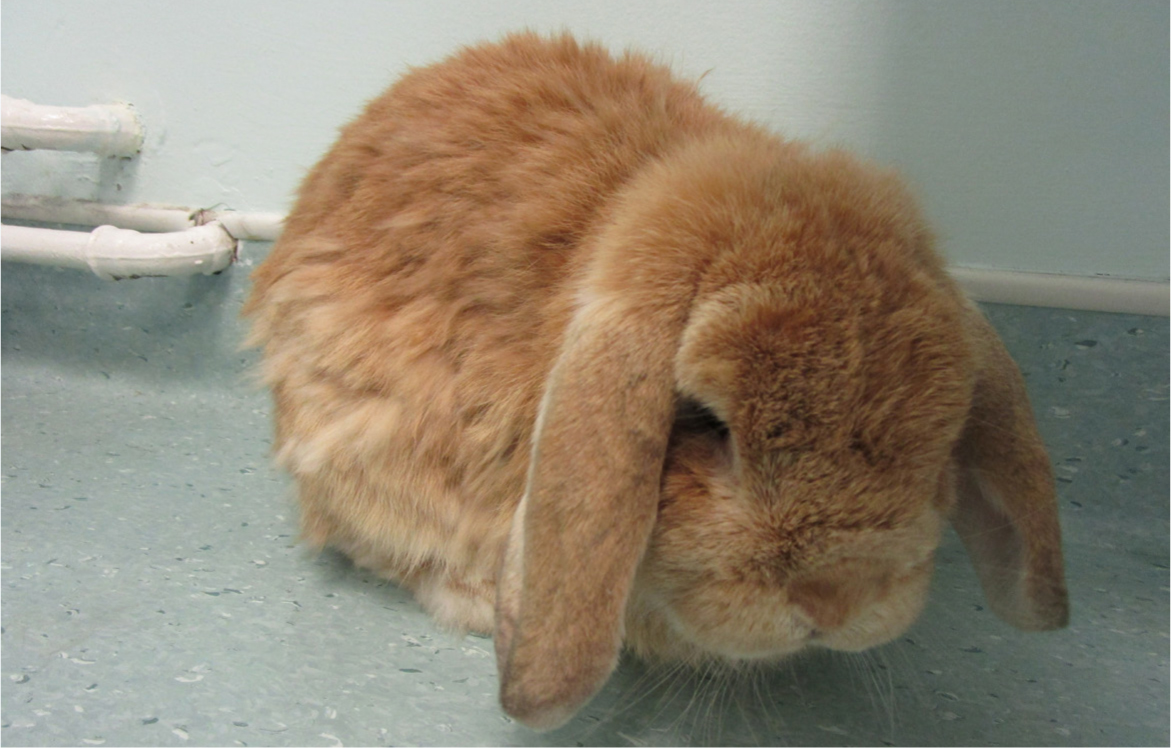
A third possibility is that even experienced veterinary personnel are not routinely examining the gait of pet rabbits on clinical examination, and are perhaps unfamiliar with subtle changes that are associated with osteoarthritis in this species. For example, pivoting on the hindlimbs or crawling gait (rather than hopping) and a hunched posture (Figure 12; Video 1). Consequently, osteoarthritis in rabbits may be difficult to diagnose clinically.
The present study had several limitations. Only rabbits attending a single centre for veterinary care were included, and consequently the results may not be transferrable to a wider population. In addition, complete data were not available for all rabbits: in particular, breed data was not sufficient for analysis. Subjective factors such as body condition and clinically apparent disease were dependent on notes recorded by the veterinarian and were subject to a level of individual variation, which is a disadvantage of retrospective studies. In addition, this study only considered variables at a single point in time and did not consider historic changes for dynamic variables such as weight and body condition, which could have impacted significantly on osteoarthritis development in the past. Further studies could be beneficial to evaluate other potential risk factors for the development of osteoarthritis in pet rabbits, such as the effect of diet, housing, substrate, exercise, comorbidities and social group interaction. Comorbidities were not investigated further, but could have influenced the development of osteoarthritis in the study population. For example, severe spondylosis of the spinal vertebrae could affect locomotion and gait, with subsequent development of osteoarthritis. Overall, further studies, in particular multicentre prospective cohort studies, are needed to make comment on the true prevalence and risk for osteoarthritis in pet rabbits, and larger sample populations are needed to evaluate the influence of breed on development of osteoarthritis.
Conclusions
This study found incidental computed tomography evidence of osteoarthritic changes in 19.6% of the sample population. Risk factors associated with these changes included greater age, greater weight and neuter status in females. Body condition score, sex and neuter status in males were not found to have a significant relationship with presence of computed tomography changes. Only 14.8% of rabbits with computed tomography evidence of osteoarthritis had clinical findings consistent with osteoarthritis in the clinical notes. This finding is concerning because it indicates that many rabbits, especially those advanced in age, may experience osteoarthritic pain, which remains undiagnosed. The consequence is that necessary analgesia is not provided, which is of major welfare concern.
The findings of this study highlight that osteoarthritis is an important disease in the pet rabbit population, and that rabbits of greater age, weight and female neutered rabbits may be at greater risk for the disease, even if pain is not clear on clinical examination. More work is needed to educate both owners and veterinary staff about the possible risk factors, presenting clinical signs and diagnostic findings of osteoarthritis in pet rabbits. Further investigation into this disease and its risk factors may be beneficial, especially when considering its potentially welfare threatening impact.
KEY POINTS
- Osteoarthritis is a disorder of articular joints which involves the deterioration of articular cartilage, osteophyte production and remodelling of bone in response to chronic damage to cartilage.
- In rabbits, while pain caused by osteoarthritis is alone a significant factor, secondary associated issues such as lack of caecotrophy, pododermatitis, cystitis and urine scald also have a detrimental impact on welfare.
- Rabbits are generally stoic and are known to hide signs of pain, and consequently, chronic issues such as osteoarthritis may go unnoticed until advanced.
- Increased understanding of risk factors for osteoarthritis in rabbits will allow preventative measures and management to be implemented against this welfare-threatening disease.
- This study concludes that rabbits of greater age and weight, and female neutered rabbits may be at increased risk of appendicular osteoarthritis. However, changes on imaging may relate poorly to clinically apparent disease.